|
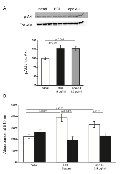
| Figure 3: HDL and apo A-I supplementation induce the phosphorylation state of Akt and increase nitric oxide in mesenchymal stromal cells in a phosphatidylinositol-3-kinase-dependent manner. (A) Representative Western blots of p-Akt and Akt in MSC 3 min after HDL (black bar) or apo A-I (grey bar) supplementation. Bar graph represents the mean ± SEM of the p-Akt/Akt ratio expressed as the percentage of the non-stimulated basal group (n=10/group for basal, HDL, and n=7 for apo A-I). (B) Bar graphs representing the mean ± SEM of intracellular NO production depicted as absorbance at 515 nm in untreated (open bars) MSC and MSC pre- and co-treated with the phosphatidyl-inositol-3-kinase inhibitor Ly 294002 (black bars) and supplemented with 5 μg/ml of HDL or 3.5 μg/ml of apo A-I for 5 min (n=10/ group). |