|
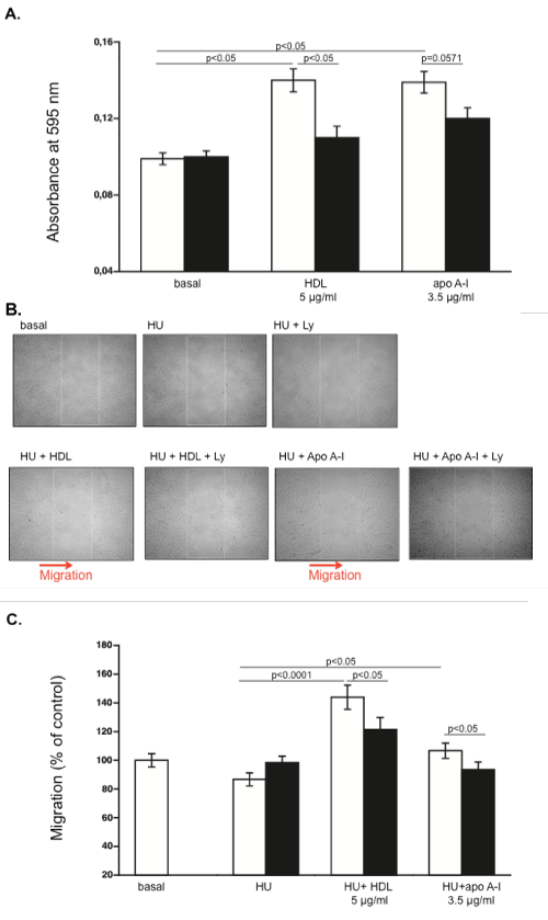
| Figure 5: HDL and apo A-I induce migration of mesenchymal stromal cells in a phosphatidyl-inositol-3-kinase-dependent manner. (A) Bar graph represents the mean ± SEM of the absorbance at 595 nm depicting the migration potential of MSC versus basal medium, HDL or apo A-I in the absence (open bars) or presence (black bars) of the phosphatidyl-inositol-3-kinase inhibitor Ly 294002 with n=4/group. (B) Representative pictures of a wound healing assay showing MSC 24 h post-scratching and supplementation of basal medium, medium containing hydroxyurea (HU) with or without HDL or apo A-I in the absence or presence of Ly 294002 (Ly) as indicated. (C) Bar graph represents the mean ± SEM of the percentage of migrating MSC 24 h after scratching and supplementation of basal medium, medium containing HU with or without HDL or apo A-I in the absence (open bars) or presence (black bars) of Ly 294002, expressed towards the percentage of the non-stimulated basal group set as 100. n=10-12 fields/group. |