|
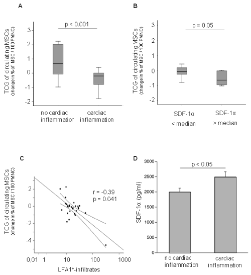
| Figure 2: (A) Quantitative analysis of transcardiac gradients of MSC in patients without (n = 6) and with (n = 23) cardiac inflammation - as defined by the number of LFA-positive cells are given as median and IQR; whiskers represent 95% CI). (B) Transcardiac gradient of MSC after dichiotomisation with respect to SDF-1α mRNA in their EMB (cut-off: median, box plots are given as median and IQR; whiskers represent 95% CI). (C) Correlation between transcardiac gradient of MSC with cardiac inflammation. (D) Bar graphs represent the mean ± SEM of post-coronary SDF-1α levels (pg/ml) in patients without and with cardiac inflammation, as indicated. |