 |
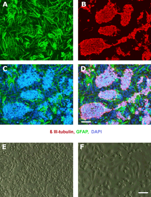
| Figure 1: Differentiating neural stem cell progeny and isolating neuronal cells. (A-D): Representative micrographs of the neuroblast assay (NBA) culture, 4 days after switching to growth factor free medium and stained for the astrocyte marker; GFAP (A), neuronal marker; β III-tubulin (B) and counterstained with DAPI (C). The colonies of β III-tubulin positive neuronal cells are on top of the astrocyte monolayer (D). (E-F): The NBA culture before (E) and after (F) shaking. After shaking the majority of the top neuronal cell clusters are detached from the underlying astrocytic cell monolayer. Scale bars = 50 μm. Abbreviations: GFAP= Glial fibrillary acidic protein, IR= Immunoreactive, DAPI= 4’,6-Diamidino-2-Phenylindole. |