 |
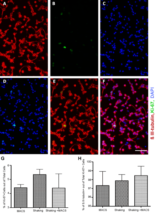
| Figure 3: Percentage of Ki-67 dividing cells in the isolated cells based on different purifying methods. (A-F): Representative micrographs of the isolated cells immunostained for neuronal marker; β III-tubulin (A), cell proliferation marker; Ki-67 (B), DAPI counterstain (C), DAPI-Ki-67 (D), β III-tubulin-Ki-67 (E), and β III-tubulin- Ki-67-DAPI merged (F). (G-H): Comparing the mean percentage of Ki-67 IR cells (G) and β III-tubulin co-expressing Ki-67 IR cells (H) in the isolated cell populations based on the MACS, Shaking and Shaking + MACS methods. As evident, the majority of Ki-67 IR cells are expressing ß III-tubulin, confirming their neuronal identity. No significant differences were detected between purifying methods. (mean ± SEM; n=3 independent experiments). Scale bars = 50 μm. Abbreviations: GFAP= Glial fibrillary acidic protein, IR= Immunoreactive, DAPI= 4’,6-Diamidino-2-Phenylindole. |