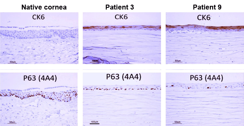
CK6: cytokeratin 6; a differentiation marker of the oral mucosa CK6 was not expressed in the native control cornea. But it was expressed in all differentiated layers of the epithelium of the post-CAOMECS trepanned corneas except in the basal proliferative layer. It showed the oral mucosa origin of the new epithelium.
P63: Antigen P63, a proliferation marker As in the native control cornea, for all the excised corneas at least one year after CAOMECS, nearly all the basal cells expressed p63; a proliferation marker more or less expressed depending on the donor, proving the regenerative capability of the CAOMECS procedure.