|
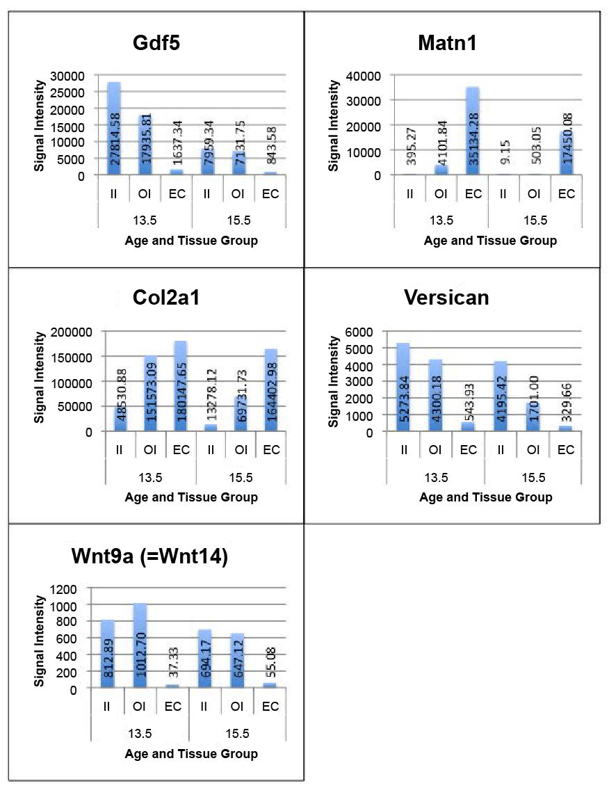
| Figure 2: Chart comparing the gene expression of Col2a1, Gdf5, Matn1, Versican and Wnt 9a (=Wnt 14) in the various sample groups. Each bar represents the average signal intensity (y-axis) of the three biological replicates of each tissue and age group (x-axis). The relative signal intensities of the three tissue types of each individual mouse embryo (biological replicate) are well exemplified by this representation of the average. |