|
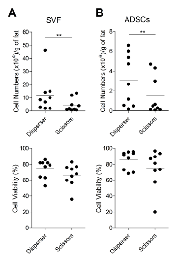
| Figure 2: Yield of SVF cells and ADSCs from the excised fat tissue (EFT) by the 2 methods. (A) Following SVF isolation, the cell numbers and viability were counted using an automated cell counter ADAM-MC with PI. (B) The isolated SVF were propagated and the resulting ADSC populations were cultured for 5 days. Cells numbers and viability were collected using an automated cell counter Vi-CELL AS with the trypan blue dye. Data were analyzed by Wilcoxon signed rank test, **p<0.01. Data show results from 9 donors and bars represent population averages. |