|
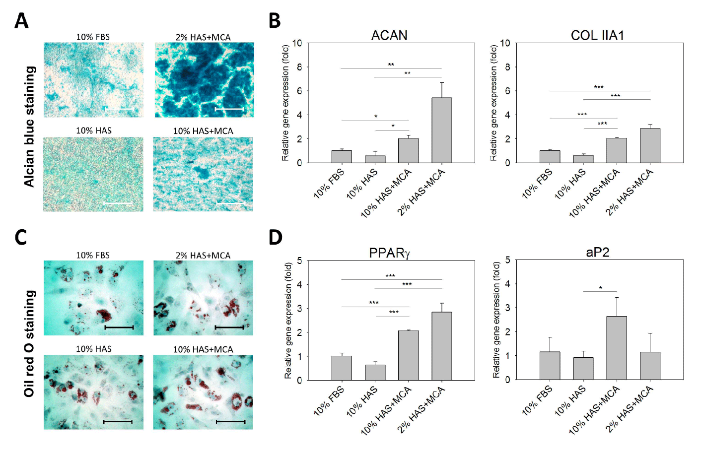
| Figure 6: Chondrogenic and Adipogenic differentiation of the ADSCs. After incubation under the different culture media for 6 days (n=3), the ADSCs were then exposed to the chondrogenesis and adipogenesis differentiation medium for 14 and 7 days, respectively. (A) Photomicrographs show sulfated proteoglycan-rich matrix (blue color) indicating chondrogenic differentiation in the ADSCs at day 14. (B) The expression of chondrogenesis-associated genes (ACAN and COL IIA1) at day 14. (C) Photomicrographs show lipid spheres (by Oil Red O staining) indicating adipogenic differentiation in the ADSCs at day 7. (D) The expression of adipogenesisassociated genes (PPARγ and aP2) in the ADSCs at day 7. For (A/C), bar=500 μm. For (B/D), β-actin was the internal control and each bar represents mean cell density ± SD (n=3). *p<0.05. **p<0.01. ***p<0.005. |