|
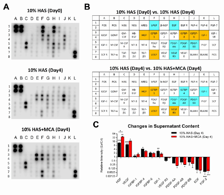
| Figure 8: The cytokine profiles of the ADSCs exposed to the different culture media (n=3). Supernatants harvested from the cultured ADSCs were compared by cytokine array on day 4. (A) The cytokine arrays. (B) Map of the cytokine arrays. Blue indicates reduction in the supernatant and orange indicates cytokine secretion. (C) Relative ratio of cytokine expression levels from cytokine array membranes. Cytokine and growth factor expression from culture supernatants were normalized against an internal positive control (POS) on the same membrane and presented as the relative intensity ratio of cultured, experimental (Ex) groups at day 4 to uncultured, control (Crl) group at day 0 (Ex/Crl). Data are presented as mean ± SD (n=3). The relative ratios of less than 1 are reduction of content in the supernatant, and the relative ratios of greater than 1 are classified as secretion. *p<0.05. **p<0.01. ***p<0.005. |