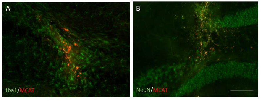
A) MCAT cells survived after the application of MES for 5 days and were detected in clusters in MCAT-MES transplanted mice;
B) MCAT cells close to the site of injection (DG), and also found scattered in the path of the needle MCAT-MES. Scale bar 100 µm.