|
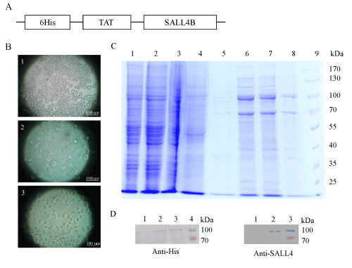
| Figure 1: Expression and purification of TAT-SALL4B protein. (A) Schematic illustration of TAT-SALL4B fusion protein construct. (B) Sf9 cell morphology at different times post-infection. 1: uninfected Sf9 cell. 2: Sf9 cells at 72 h post-infection. 3: Sf9 cells at 120 h post-infection. (C) Purified TATSALL4B was resolved on a 10% denaturing gel and stained with Coommasie brilliant blue. Lane 1: lysates of uninfected Sf9 cells. Lane 2: lysates of infected Sf9 cells. Lane 3: flow through fraction. Lane 4 and 5: wash fractions. Lanes 6, 7, 8: elution fractions 1, 2, 3. Lane 9: molecular markers. (D) Western blot analysis with anti-His antibody. Lane 1: uninfected Sf9 cells. Lane 2, 3: Infected Sf9 cells. Lane 4: molecular markers. (E) Western blot analysis with anti-SALL4 antibody. Lane 1: uninfected Sf9 cells. Lane 2: Infected Sf9 cells. Lane 3: molecular markers. |