|
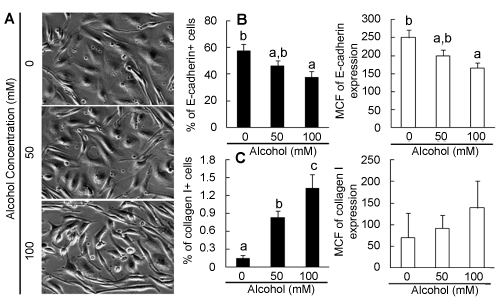
| Figure 6: A: Morphological changes of primary HL-1-1 cells. Cells were cultured in the differentiation medium without and with different concentrations of alcohol for 10 days. The images captured on the Olympus IX81 Imaging System with 10X phase objective lens represent 5 sets of cell cultures. B: Changes in E-cadherin expression by primary HL1-1 cells. Cells were cultured in the differentiation medium without and with different concentrations of alcohol for 2 weeks. N=4 sets of cell cultures. C: Changes in collagen I expression by primary HL1-1 cells. Cells were cultured in the differentiation medium without and with different concentrations of alcohol for 2 weeks. N=4 sets of cell cultures. Values are mean ± SEM. MCF=mean channel fluorescence. Bars with different letters in each panel are statistically different (p<0.05). |