 |
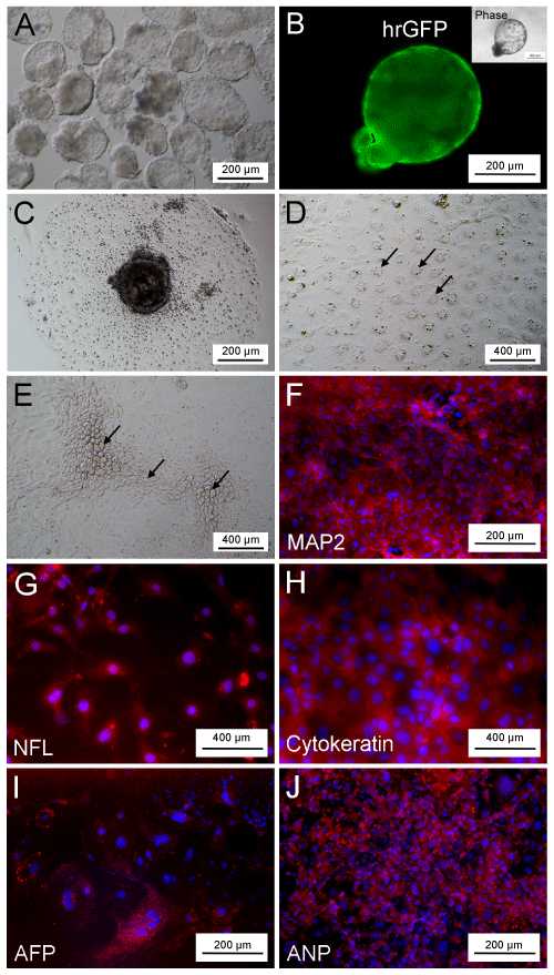
| Figure 4: In vitro vitro embryoid body formation and differentiation of piPS/ hrGFP+ cells. (A) EB formation of piPS/hrGFP+ cells on day 7 after hanging drop culture. (B) hrGFP expression of EB. (C) EB expansion after attachment to gelatin-coated surface of culture dishes. (D) The nissl Nissl bodies of the cells derived from attached EB (Black arrows). (E) The epithelial cells of cells derived from attached EB (Black arrows). Immunocytochemistry staining of cells derived from attached EB to antibodies against (F) MAP2 (F), (G) NFL (G), (H) Cytokeratin (H), (I) AFP (I), and (J) ANP (J) antibodies. Nuclei were stained with DAPI (blue). |