 |
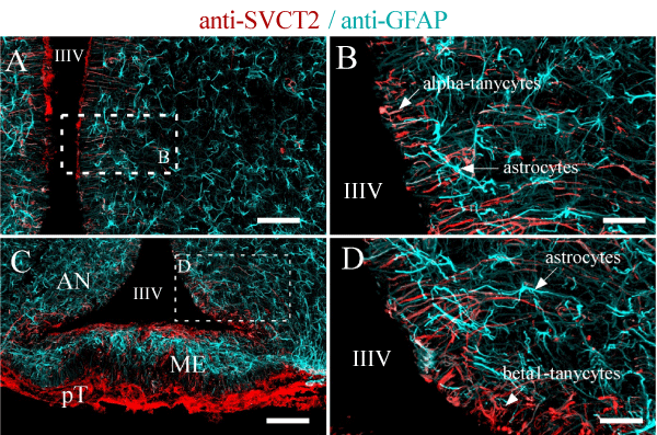
| Figure 4: SVCT2 is highly expressed in hypothalamic tanycytes. Tanycytes and astrocytes are specialized glial cells distributed in the hypothalamus. Tanycytes can be classified as alpha (A-B) or beta (C-D), which contact the CSF of the third ventricle (apical area); the processes contact different areas of the hypothalamic region (basal area). Astrocytes are present in the hypothalamic sub-ependymal region and are concentrated in the median eminence (C, light-blue staining) below the ventricular layer (beta-2 tanycytes). SVCT2 and GFAP immunofluorescence analysis was performed using confocal spectral microscopy (Zeiss 780 equipment), tile scanning and Z-stack projection imaging. An intense immunoreaction for SVCT2 was detected in alpha and beta tanycytes (red signal); however, astrocytes, endothelial cells, and neurons were negative for SVCT2 staining. Astrocytes showed an intense immunoreaction for GFAP (light-blue staining). IIIV, Third ventricle; AN, Arcuate nucleus; GFAP, glial fibrillary acidic protein; ME, Median eminence; pT, Pars tuberalis. Scale bars in A and C, 500 μm; in B and D, 40 μm. |