 |
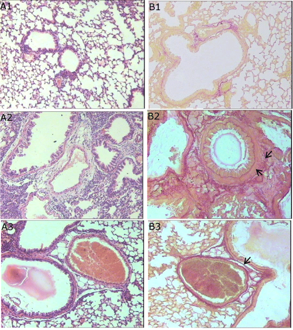
| Figure 1: Photomicrographs of representative lung sections obtained from C57BL/6 mice. (A) Tissues were stained with haematoxylin and eosin in order to investigate inflammatory cells accumulation (7th day of the experiment); (B) tissues were stained with picrofuchsin to determine the collagen content (21st day of the experiment). (A1, B1) Mice receiving intratracheal NaCl 0.9%, (A2, B2) Mice receiving intratracheal bleomycin, (A3, B3) Mice with fibrosis treated spiperone. A, pink-purple stains indicate cytoplasm, and blue the nuclei of inflammatory cells. A2, on the 7th day after bleomycin instillation it was observed the infiltration of alveolar and alveolar ducts interstitium by lymphocytes, macrophages, neutrophils, plasmocytes. A3, spiperone reduces the degree of alveocyte desquamation in alveolar lumen. B, dark pink stains are collagenous deposits. B2, the most expressed collagen fibers deposition after bleomycin injection was observed on the 21st day. B3, it is shown decrease in area of collagen deposition in spiperone-treated mice. The photomicrographs were taken using an Axio Lab.A1 (Carl Zeiss MicroImaging GmbH; Göttingen, Germany) microscope and AxioCam ERc5s digital camera. All photomicrographs were at 100 × magnification. |