|
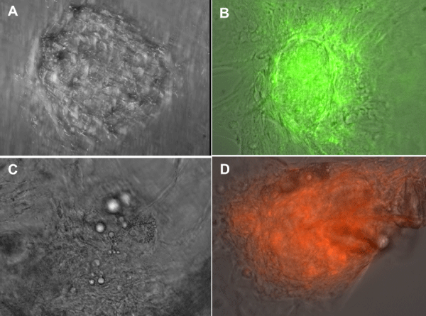
| Figure 4: MCs growing on Matrigel incubated with LCDD and amyloidogenic LCs for 4 days. A,B-Phase contrast microscopy. AX750; BX750; C, D-Mesenchymal stem cells marked with fluorescent PKH green (C) and D with Magic Red. CX750, DX750. MCs incubated with LCDD-LC form mesangial nodules while MCs incubated with amyloidogenic LCs engage in the formation of amyloid. A,B-MSCs migrate to sites of damage (C,D). |