 |
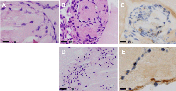
| Figure 9: MCs growing on Matrigel platform incubated with amyloidogenic and LCDD LCs followed by introduction of RMSCs. A-C- Hematoxylin and eosin, X500, D- Hematoxylin and eosin, X350; E- Immunohistochemical stain for smooth muscle actin, X350. MCs incubated with amyloidogenic LC formed amyloid (A) and with LCDD-LCs formed nodules with excess extracellular matrix (B,C). Interaction of MCs with MSCs is clearly shown in D. Apoptotic MCs are eventually replaced by MSCs which differentiate into MCs expressing muscle specific actin in their cytoplasm (E). |