|
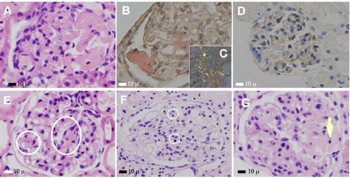
| Figure 11: Ex-vivo kidney perfusion platform. Injection of amyloidogenic (A-D) and LCDD (E)-LCs via renal artery followed by injection of MSCs (F,G) through renal artery. AX350, Hematoxylin and eosin stain; BX350, Congo red stain; CX500, polarization of Congo red stained section; DX350, immunohistochemistry stain for smooth muscle actin; EX350- Hematoxylin and eosin stain; FX350- Hematoxylin and eosin stain; G-X350- Hematoxylin and eosin stain. Amyloid formation with Congo red staining and apple green birefringence occurring when MCs were incubated with amyloidogenic LCs (A-D) and expanded mesangial areas with extracellular matrix forming small mesangial nodules (E). MSCs in capillary lumina in glomerulus (F) (circles) and migrating to site where amyloid formation (arrow) has occurred in a glomerulus (G) to participate in the repair of the damaged mesangium. |