|
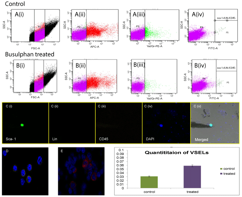
| Figure 4: Quantitation of VSELs: Quantitation of VSELs in (A) control and (B) busulphan treated mice testes by flow cytometry. Cells between 2-6 μm were gated using size calibration beads (Ai,Bi) followed by sequential selection of LIN negative population (Aii,Bii), CD45 negative population(Aiii,Biii) and then SCA-1 positive population in both control (Aiv) and busulphan treated (Biv) testes. Confocal images of sorted VSELs show that SCA-1 positive cells were negative for LIN and CD45 (Ci-v). Specificity of APC tagged LIN (D) and PE tagged CD45 (E) antibodies analyzed using mouse bone marrow cell smears as positive control. (F) shows percentage of VSELs obtained in testicular cell suspension prepared from control and busulphan treated mice. Error bars represent standard deviation between three biological replicates. |