|
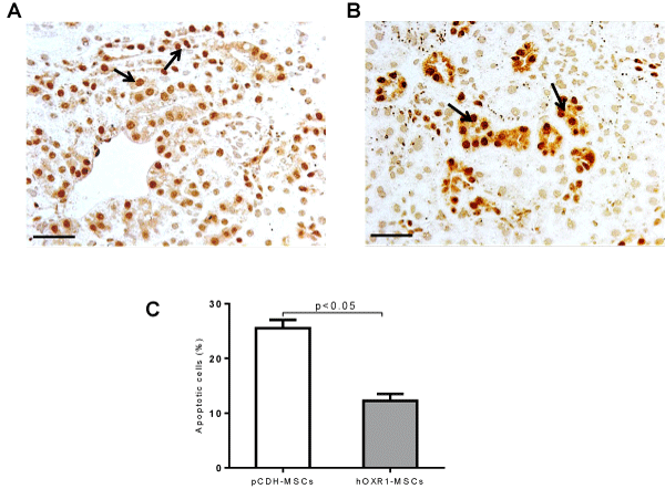
| Figure 7: Tunel staining for apoptosis detection in B6.Sle1.Sle2.Sle3 kidney. (A-C) Detection of apoptotic cells in the kidneys of B6.Sle1.Sle2.Sle3 mice treated with pCDH-MSCs (A) or hOXR1-MSCs (B) for 8 weeks. Apoptotic cells appeared dark brown (indicated by arrow). (C) Plotted is the quantitative analysis of the percentage of renal apoptotic cells in the 2 groups of mice. (Original magnification 400X, scale bars=50 μm). Each group had 5 mice. |