|
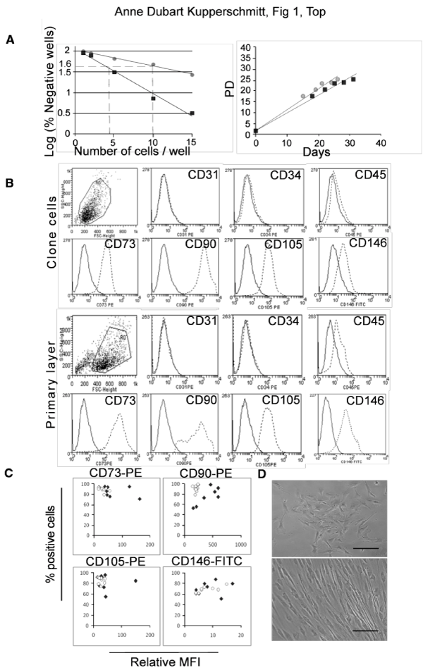
| Figure 1: Clone Generation and Phenotypic Characterization A) Clones generation from primary layers seeded at limiting dilutions when first passaged. Left: Limiting dilution. The cloning efficiency, calculated after verification of the Poisson distribution of the clonogenic cells, was 1 in 4 and 1 in 10 (r2=0.99) in the two representative experiments shown. Right: Cumulative population doubling (PD) of two representative clones. Linear adjustment (r2=0.98). Day 0 indicate the day of limiting dilution corresponding to the first passage of the primary layers. B) Flow cytometry analysis of mesenchymal (CD73, CD90, CD105, CD146), endothelial (CD31, CD34) and hematopoietic (CD45) markers for one representative clone (clone 4 from fetal liver 3: c4FL3) compared to its corresponding primary layer. C) Relative Mean Fluorescence Intensity (MFI) vs. the percentage of positive cells for the different mesenchymal markers. Results for primary layers (n=9, white circles) and clones (n=7, black diamonds). MFI for each antigen was related to MFI of a corresponding isotype control. D) Phase-contrast microscopy photographs showing the adherent cell morphology of a representative clone (c4FL3). Upper: non confluent culture (5 days after seeding); lower: confluent culture (14 days after seeding). Bar: 50 µm. |