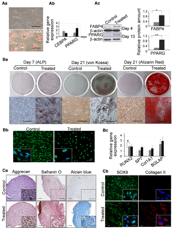
A) Adipocytic
a) Morphology of c4FL3 cells stained by Oil Red. Upper: Control culture (untreated) grown in expansion medium for 30 days. Lower: Cells cultured in adipocytic conditions (treated) for 30 days; Bar: 20µm
b) Q-RT-PCR data showing the expression of PPARG and CEBPA in 3 clones (c4FL3, c3FL8, c7FL8) at day 4 and day 21 after the induction of differentiation. Values are expressed as mean ± SEM of gene expression normalized to that of GAPDH and related to that at day 0 (ΔΔCt method). White bars: day 0; gray bars: day 4; black bars: day 21. Values at different time-points are not significantly different.
c) Western blot analysis of fatty acid-binding protein 4 (FABP4) and peroxisome proliferator-activated receptor gamma (PPARG) protein levels in clones at day 4 and day 10 after adipocytic induction, respectively. Left: representative western blot (c4FL3). Right: densitometric studies. Mean ± SEM of values obtained for clones c4FL3, c3FL8 and c7FL8. Value for each protein was related to that of β-actin. Gray bars: protein extracts of non-treated cells; black bars: protein extracts of cells 4 or 10 days after adipocytic induction. *p <0.05, **p<0.01.
B) Osteoblastic a) Histochemistry for clone c4FL3. Left: alkaline phosphatase (ALP) staining showing the initiation of osteoblastic differentiation at day 7 after induction. Centre and Right: mineralization assessed by von Kossa and alizarin stains at day 21. Bars: 50µm. b) Immunofluorescence using antibodies against transcription factor RUNX2 for clone c4FL3 in non-induced (untreated) cells or at day 4 after induction of osteoblastic differentiation (treated). Bars: 10µm. c) Q-RT-PCR data showing expression of RUNX2, SP7, Col1A1 and BGLAP in 3 clones (c4FL3, c3FL8 and c7FL8) at day 4 and day 14 after differentiation induction. Values are expressed as mean ± SEM of gene expression normalized to that of GAPDH and related to that at day 0 (ΔΔCt method). White bars: day 0; gray bars: day 4; black bars: day 14. *p<0.05.
C. Chondrocytic a) Aggrecan, safranin O and alcian blue stainings for clone c4FL3 at day 21 in absence of induction (untreated) or after differentiation induction (treated). For each staining, a magnification view is shown in insert. Bars: 25 µm. b) Immunofluorescence for transcription factor SOX9 and for collagen II in the representative clone (c4FL3) in absence of induction (untreated) or at day 7 after differentiation induction (treated). Bars: 10 µm.