 |
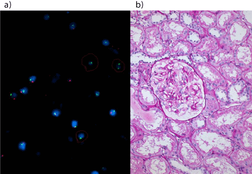
| Figure 4 a) On left, peripheral blood lympho-hematopoietic chimerism demonstrated by fluorescent in situ hybridization of the same patient performed on the day of protocol biopsy (cells with 2 green dots depict female cell with XX chromosomes highlighted by circle, against cells with green and red dots depicting male cell with X and Y chromosomes). b On right, Normal protocol biopsy of patient belonging to group-2; 44 years old man underwent renal transplantation for chronic renal failure due to unexplained etiology, with 38 years old wife’s kidney (HLA 0/6 match) on 17th August, 2012, biopsy date 2nd August, 2013, immunosuppression: Sirolimus, 1 mg/ day and Prednisone, 5 mg/day. Periodic Acid Schiff stain, x 200. |