|
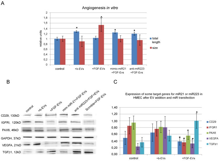
| Figure 3: Effect of HMEC transfection with mimic miR-21 or with anti-miR-223. (a) Diagram of vessel-like structures formed in vitro by non-stimulated HMEC (control), by HMEC stimulated with b-EVs or FGF-EVs, or by transfected HMEC stimulated with FGF-EVs. Total length (blue column) and size (red column) of vessel-like structures was measured, (mean ±SEM, * - p<0,05 vs. “control total length”, ^ - p<0,05 vs. “control size”, n=15); (b) representative Western blot of proteins, that are targets of miR-21 or miR223; (c) expression of miR-223 or miR-21 target proteins after EV addition (data are represented as densitometric evaluation of band mean intensity ±SEM, * - p<0,05 vs. “+b-EVs”, n=5).. |