 |
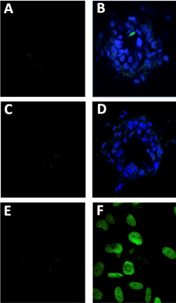
| Figure 3: Immunohistochemical assays of stem cell biomarker expression in adult spinal disc stem cells. Discospheres and single stem cells were resuspended in stem cell media, plated on laminin-coated cover slips, incubated for 8 hours to allow attachment, and then fixed and processed for biomarker expression studies. A. C. E. Negative controls for immunostaining of CD133 (A), Nestin (C), and Survivin (E). B. D. F. Immunohistochemistry for the expression of these stem cell biomarkers. B and D. The expression of CD133 (B) and Nestin (D) in discospheres attached to laminin coated coverslips for 8 hours. Photomicrographs taken with fluorescent confocal microscopy at 40X magnification demonstrate uniform and high expression of both biomarkers throughout the sphere structure. F. The expression of Survivin in adult spinal disc stem cells plated on laminin coated culture surfaces as single cell suspensions, as shown in photomicrographs taken with fluorescent confocal microscopy at 40X magnification, demonstrate uniform and high expression of Survivin biomarkers in attached spinal disc stem cells. |