|
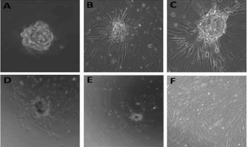
| Figure 7: 2-Dimensional Tissue engineering with 3-dimensional discospheres (the burst kinetic assay) Discospheres were plated on coverslips coated with 0.1% gelatin, and cultured in chondrogenic media and culture conditions for 5 days. Imaging data was collected at 24 h intervals with inverted light microscopy at various magnifications, starting on day 1, ending on day 5. A. Photomicrograph taken with light microscopy at 60X magnification of a discosphere cultured in the burst kinetic tissue-engineering assay at 24 hours. B. and C. Photomicrographs taken with light microscopy at 60X magnification of a discosphere cultured in the burst kinetic tissue-engineering assay at 48 hours (B) and 72 hours (C) demonstrating a dramatic “blastic” phase. As shown in the figure, the cells at the sphere edge change dramatically in phenotype, and become elongated spindle shaped cells that migrate from the sphere and change phenotype and morphology. D. and E. Photomicrographs taken with light microscopy at 20X magnification at 96 hours (D) and 120 hours (E) of a discosphere cultured in the burst kinetic tissue engineering assay; demonstrating the appearance of small rounded cells that migrate out across the culture surface away from the sphere, and gain and cell migration and proliferation biology. F. Cultures allowed to continue to grow were followed over time. Long-term studies with these assays demonstrated that these cells continue to migrate and proliferate, until the culture surface was crowded with cells. At this time, the NP cells change morphology and phenotype, differentiated into mature NP cells, ceased migration and proliferation, and secreted significant extracellular matrix. |