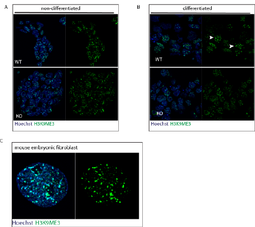
A. H3K9ME3 immunofluorescent staining on non-differentiated WT and KO ESC.
B. H3K9ME3 immunofluorescent staining on 7 days differentiated WT and KO ESCs. Note the H3K9ME3 condense staining in the different WT cells, as indicated with white arrows which is absent in the Dgcr8 KO ESCs.
C. H3K9ME3 immunofluorescent staining on mouse embryonic fibroblast served as a positive control.