|
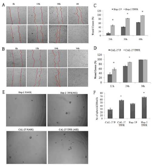
| Figure 3: PFR cells showed increased migratory and self renewal properties(A&B). The migratory ability ofHep2-TPFR and CAL-27 TPFR cells was assessed by the wound healing assay. (A) The closure of Hep-2 parental cells (Upper panel) and TPFR (Lower panel) was compared at different time points of 0, 24, 30 and 48 h. At 48 h, Hep-2 TPFR completely closed the wound as compared to 64.14% of closure by its parental cells. (B) The closure by CAL-27, and the TPFR was documented from 0-36 h. CAL-27 TPFR and parental cells closed the wound by 36 h but the percentage wound closure by CAL-TPFR at 12h and 24 hr was higher than that of its parental cells (Magnification is 200X). Graphical representation of the percentage of wound closure (SEM±SE, three independent experiments, p≤.05) at different time points is also shown (C & D). The number of spheroids (40X) generated by Hep-2 TPFR cells (upper panel) and CAL-27 TPFR (Lower panel) in low attachment serum free condition was more that their parental cells (E). Graphical representation of the results are shown (F) as SEM±SE of three independent experiments. |