|
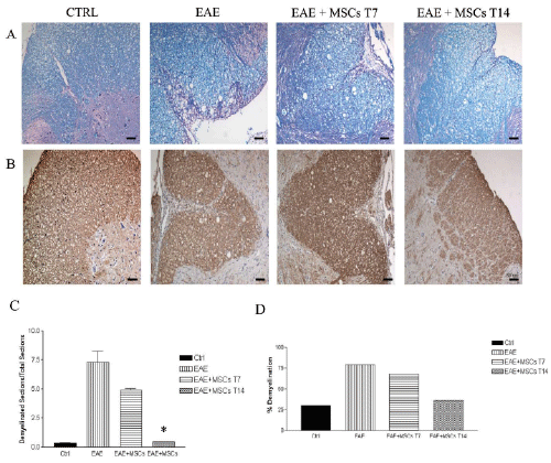
| Figure 3: Histopathological examination of demyelination in RR-EAE rats.Representative sections were obtained from lumbar spinal cords of untreated CTRL rats, EAE rats, and EAE rats treated with MSCs injected after 7 (T7) or 14 days after the immunization (T14). Sections were stained with Luxol Fast Blue and eosin (A), or immunostained with anti- MBP and hematoxilin (B). Scale Bar 50μm. Arrow indicated demyelinated area, which resulted unstained by Luxol Fast Blue.C) The number of sections presenting demyelination on the full amount of sections was then analyzed, as well as the percentage of demyelinated lesions for each samples (D). * P< 0.05, EAE+MSC T14 vs. EAE. |