|
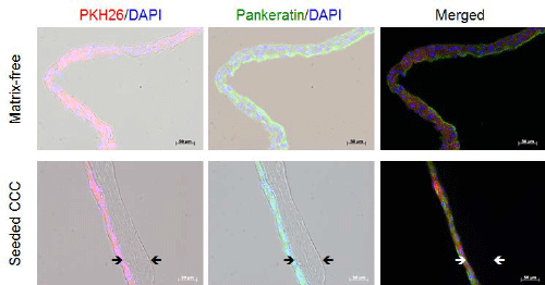
| Figure 5: Immunofluorescence analysis of epithelial phenotype. For characterising stratified cultures of PKH26-labelled HUC seeded in high-density on CCC the expression of pankeratin, a marker for epithelial phenotype, was determined via immunofluorescence technique. With reference to detached matrix-free controls from standard plastic culture (top row) a comparable homogeneous staining pattern of pankeratin could be detected for in vitro generated urothelium on CCC (bottom row). Red fluorescence for PKH26 and green fluorescence for pankeratin were shown together with blue nucleus signals from DAPI in bright field (left/centre) or as merged (right) photographs. |