 |
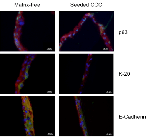
| Figure 6: Detection of urothelial marker expression. With reference to detached matrix-free controls from standard plastic culture (left column) in vitro generated urothelium of PKH26-labelled HUC seeded in high-density on CCC (right column) showed comparable expression pattern of p63, K-20 (both: ongoing differentiation), E-cadherin and ZO-1 (both: junction formation). Red for PKH26, green for the specific marker and blue signals from DAPI were shown as merged photographs. |