 |
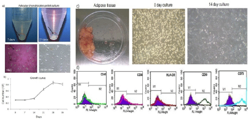
| Figure 1: Cell morphology and characterization of articular chondrocytes and hADSCs. (a) Formation of chondrocyte micro mass aggregates after 7 and 21 days of pellet culture. H&E staining shows polygonal shape of chondrocytes in isogenous group with lacunae and alcian blue positive staining indicating glycosaminoglycans abundantly distributed throughout the cells. (b) Growth rate of chondrocytes with increased proliferation after 21 days (n=5). There was no increase in the cell number after 7 days of culture. The cell number increased to 8 × 10⁴ cells/mL on 28 day and started to decline after 30 days. (c) hADSCs isolated from adipose tissue by enzymatic digestion. Cells attained spindle shaped morphology on 14 day. (d) The fluorescence level detected for the stromal cell markers was 98.5% (CD90), 90% (CD73), whereas for CD34/45, HLA-DR was observed only 1-3% of the gated cells. The positive zones in the histograms were marked as M2 gate based on the control sample. |