 |
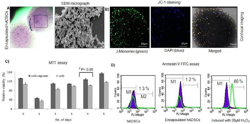
| Figure 2: Morphological evaluation of alginate encapsulated hADSCs. (a) The alginate-hADSCs suspension was added drop wise on CaCl2 solution and incubated for 15 min at 37°C in a humidified atmosphere of 5% CO2. The giemsa staining of encapsulated hADSCs showing uniform distribution of living cells within alginate bead. SEM micrographs of cross sectioned encapsulated cells showed distribution of hADSCs on the matrix and exhibited a round-shaped morphology due to the lack of interactions between cells and alginate as shown by an arrow. (b) JC-1 dimer formation within the alginate bead, thereby indicating the presence of metabolically active live cells on the hydrogel using CLSM. The monomer emitting at 527 nm (green) after excitation at 490 nm, and J-aggregates emits at 590 nm (orange dots) demonstrating the viability of cells. The DAPI counter staining shows the presence of stem cells within in the alginate matrix. (c) A statistically significant (p< 0.0.5) differences was observed for the cells cultured within the alginate matrix up to 7 days as evident from MTT assay (n=6). The viability was observed >90% after 4 days in the encapsulated cells. (d) Annexin-V-FITC of encapsulated cells did not undergo apoptosis when cultured within the alginate matrix. M1: region of fluorescent cells with intact membranes (living cells) and M2: region of non-fluorescent cells with damaged cell membranes (dead cells).In contrast, cells induced with H2O2 underwent apoptosis shown as shift in the peak as compared with unstained control (shown as Green overlay in the histogram. |