|
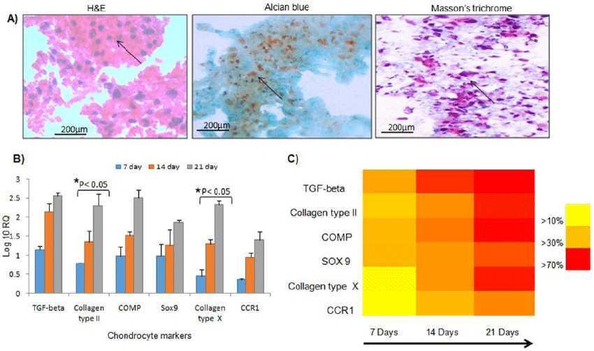
| Figure 3: Differentiation potential of alginate encapsulated cells. (a) hADSCs were cultured in alginate bead based pellet system. Hematoxylin and eosin staining of the pellets shows rounded cells within the beads; alcian blue stains the collagen matrix with more number of cells in the center as indicated by an arrow. Metachromatic staining was observed with masson’s trichrome indicating differentiated cells in red colour and suggesting presence of cartilaginous matrix. (b) Chondrogenic differentiation potential of the hADSCs within the alginate matrix was evaluated by gene expression studies using RT-PCR. The graph indicates the expression levels of various chondrogenic genes on different days. The data revealed significantly high levels of collagen type X, TGF-beta and SOX9expressions. The expression levels of collagen type II and COMP increased by 14 days of culture and reached peak level by 21 days. Data is expressed as mean ± SD with p< 0.05 (n=3). (c) Heat map analysis summarizes the expression pattern of chondrocyte specific genes of differentiated hADSCs in 3D alginate bead. The level of gene expression levels was plotted in yellow to red scale with yellow being lightest 0–20% and darkest being 50-70% level. The result depicts varied patterns of gene expression over the 21-days of the culture. The representation enables study of temporal progression of chondrogenesis. |