|
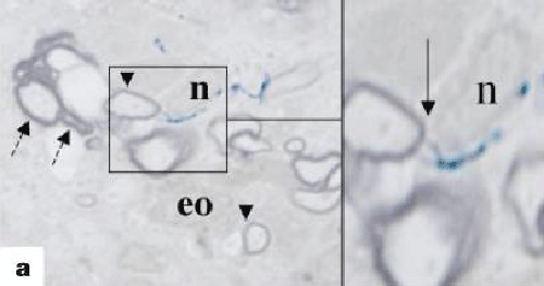
| Figure 5: Light microscopy of transplanted neural stem cells. Light microscopy showing transplanted neural stem cells contributing to myelination of axons. ‘n’ indicates the nucleus of the neural stem cells, with blue deposits going to surround the axon (arrows) surrounded by myelin (dashed arrows). The short arrowheads represent thin layer of myelin [68]. |