 |
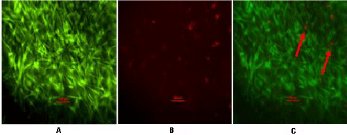
| Figure 4: Cell viability of eBMMSCs after 6 days in culture. Cells were stained with calcein-AM which exhibits green fluorescence and demonstrates live cells (A) and propidium iodide which displays red fluorescence and demonstrates dead cells (B) (shown with red arrows). Panel C represents the merged image of A and B. Fluorescent micrographs showed that in presence of PRP, eBMMSCs adhered to, were viable and proliferated in culture. Scale bar=100 μm. |