 |
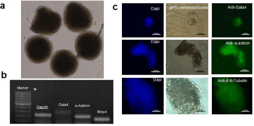
| Figure 3: In vitro differentiation (a) Embryoid body generated after 3 days of giPSCs culture in low attachment dishes, which supports pluripotent nature of giPSCs. (b) Presence of different germ layer markers GATA4, α-Actinin and BMP4 in RTPCR assay and (c) Gata4, α-Actinin and β-III-Tubulin in immunostaining confirms the presence of endodermal, mesodermal and ectodermal cells in the embryoid bodies. One scale bar is 100 μm. |