|
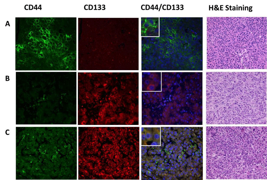
| Figure 1: Illustration of liver cancer stem cells immunofluorescence microscopy and H&E staining of explanted livers. A. Sixty year old male diagnosed with a solitary stage II HCC (CD44+), AFP level 4.8 mg/dl, recurrence-free survival of 128 months. H&E staining demonstrate a moderate to poorly differentiated tumor. B. Fortyeight year old male diagnosed with multiple stage II HCC (CD133+), AFP level 2.5 mg/dl recurrence-free survival of 93 months. H&E staining demonstrate a welldifferentiated tumor. C. Fifty five year old male diagnosed with a solitary stage I HCC (CD44+/CD133+), AFP level 3.6 mg/dl, recurrence-free survival of 39 months. H&E staining demonstrate a moderate to poorly differentiated tumor. Expression of stem cell markers was reported as single CD44 positive, CD133 positive or dual pattern of immunofluorescence positivity when expression of both markers was identified in the same cell. Magnified are represents tumor cells with positive LCSC markers. H&E magnification of 20X. |