|
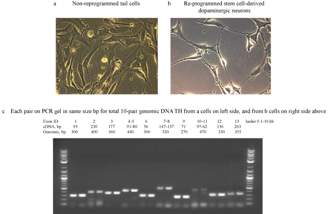
| Figure 4: Long-term cell culture. After 6 months, the neuronal cells were displaying further signs of mature network formation from the re-programmed stem cellderived dopaminergic neurons (Figure 4b). Importantly, control tail cells that were not subject to the re-programming procedures kept their original shape in the same time period (Figure 4a). Genomic DNA TH was abstracted (Figure 4c) from both cell lines (Figure 4a and 4b) for further cancer risk assessments. A total of ten pairs with the same size (bp) of PCR products (from Figure 4a and 4b) were selected to run on gel representing complete Exons 1-13 (ID numbers) of TH genomic DNA (Figure 4c). In each of the same size pairs on gel, the cells band (PCR products) on the left side were from the original control non-programmed tail cells (Figure 4a), while the cells band on right side came from the re-programmed stem cell-derived dopaminergic neurons (Figure 4b). Here, the sizes (bp) of PCR product bands on the gel showed that of genomic DNA TH in Fig. 4-c. The relevant sizes with Exons 1-13 and cDNA, which matched with related genomic DNA, were also suggested in Figure 4c in order to highlight the relationships among them. |