|
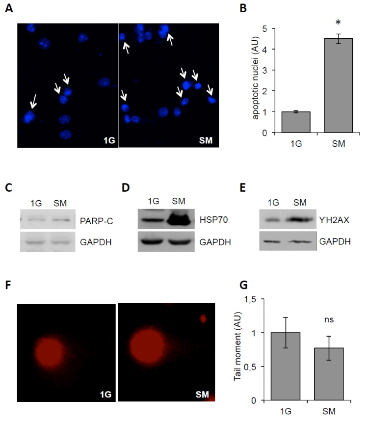
| Figure 3: SM causes apoptosis and cellular stress in NSC. (A) Representative images of pyknotic nuclei (indicated by arrows) of cells grown under SM and at 1G as control stained with DAPI. (B) Histogram showing quantification of manual counts of pyknotic nuclei in NSC grown in SM normalized against 1G as control; p<0.05. AU: arbitrary units (C) Western blot showing cleaved PARP (PARP-C) levels and GAPDH as loading control in NSC grown in SM and 1G. (D) Western blot showing HSP70 levels and GAPDH as loading control in NSC grown in SM and 1G. (E) Western blot showing active phosphorylated histone H2AX (ƳH2AX) and GAPDH as loading control in NSC grown in SM and 1G. (F) Representative images of comet assay performed on NSC grown in SM and 1G. (G) Histogram showing normalized tail moment of comet assay performed on NSC grown in SM and 1G, ns: not significant. All experiments were performed in triplicate. |