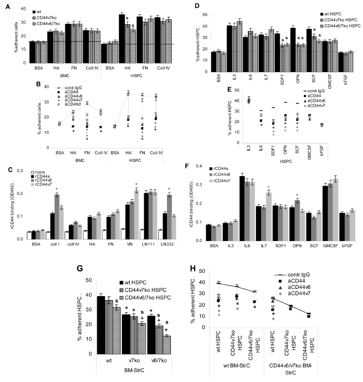
Figure 2: BMC, HSPC and BM-StrC adhesion: (A) CFSE-labeled BMC and HSPC from wt, CD44v7ko and CD44v6/v7ko mice were seeded on BSA, HA, FN or coll
IV coated plates; (B) wt BMC and HSPC were preincubated with anti-panCD44, -CD44v6, -CD44v7 and -CD49d and were seeded on BSA, HA, FN or coll IV coated
plates; (C) rCD44, rCD44v6 and rCD44v7 were seeded on BSA, HA, FN, coll I, coll IV, LN111 and LN332 coated plates; (D) wt, CD44v6/v7ko and CD44v7ko HSPC
were seeded on cytokine or chemokine-coated plates; (E) wt HSPC were preincubated with anti-panCD44, -CD44v6 and -CD44v7 and were seeded on cytokine or
chemokine-coated plates; (F) rCD44, rCD44v6 and rCD44v7 were seeded on cytokine or chemokine-coated plates; (G) CFSE-labeled wt, CD44v6/v7ko and CD44v7ko
HSPC were seeded on a monolayer of wt, CD44v6/v7ko and CD44v7ko BM-StrC; (H) CFSE-labeled wt, CD44v6/v7ko and CD44v7ko HSPC were preincubated with
anti-pan-CD44, -CD44v6 or -CD44v7 and seeded on a monolayer of wt or CD44v7ko BM-StrC (A-H) Adhesion was evaluated after 4h incubation at 37oC, 5%CO2;
(A,B,D,E,G,H) after washing, adherent cells were lysed and fluorescence was evaluated in a fluorescence ELISA reader. Adhesion is presented as % of input cells;
(C,F) Plates were washed, incubated with anti-CD44-AP and substrate. OD was determined at 450nm. All assays were performed in triplicates, mean ± SD are
presented; (A,D,G): significant differences between wt and CD44v7ko or CD44v6/v7ko cells: s; (B,E,H) significant inhibition by anti-CD44: *, significant inhibition by anti-
CD49d: +; (C,F) significant differences between rCD44s versus rCD44v6 or rCD44v7 binding: +; (G) significant differences in HSPC binding to wt versus CD44v7ko or
CD44v6/v7ko BM-StrC: +.
HSPC CD44 contributes to HA, FN and less pronounced, coll IV adhesion; CD44v6/CD44v7 strengthen HA and FN adhesion. CD44 also binds cytokines / chemokines;
CD44v6 and CD44v7 contribute to SDF1, OPN and SCF binding. Importantly, BM-StrC CD44v6/v7 also contributes to HSPC binding. |
