|
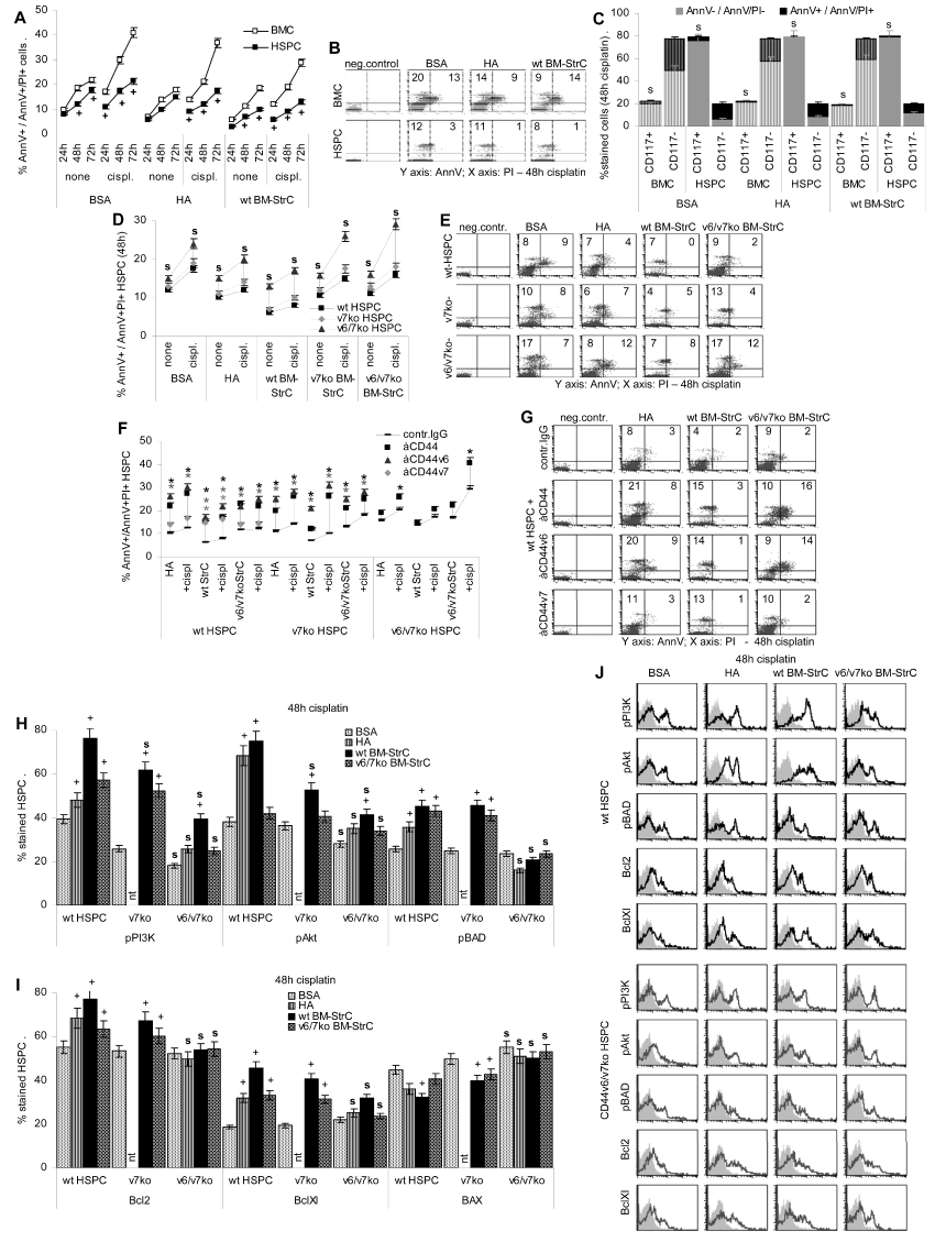
| Figure 6: Apoptosis resistance of HSPC is supported by HA and BM-StrC: (A-C) Wt BMC and HSPC and (D-G) wt, CD44v7ko and CD44v6/7ko HSPC were cultured for 24h-72h in the absence or presence of 5μg/ml cisplatin on BSA, HA or BM-StrC. Where indicated cultures contained anti-CD44; apoptosis was evaluated by AnnV/PI staining; (A,B) Comparison of the % AnnV+/AnnV+/PI+ cells in BMC versus HSPC; the mean percentage (±SD) (triplicates) of AnnV+/AnnV+/PI+ cells and representative examples are shown; significant differences between BMC and HSPC: +; (C) repetition of the experiment shown in (A) and triple staining for CD117, AnnV and PI; the mean percentage (±SD) (triplicates) of CD117+ and CD117- cells that are AnnV+/PI+ or AnnV-/PI- are shown; significant differences in AnnV+/PI+ CD117+ versus CD117- cells: s; (D,E) wt, CD44v7ko and CD44v6/v7ko HSPC were cultured in the presence or absence of cisplatin on BSA, HA or BM-StrC coated plates; the mean percentage (±SD) (triplicates) of AnnV+/AnnV+/PI+ cells and representative examples are shown; significant differences between wt, CD44v7ko and CD44v6/v7ko HSPC: s; (F,G) wt, CD44v7ko and CD44v6/v7ko HSPC were cultured in the presence or absence of cisplatin on BSA, HA or BM-StrC coated plates; the cultures contained control IgG or anti-panCD44, -CD44v6 or -CD44v7; the mean percentage (±SD) (triplicates) of AnnV+/AnnV+/PI+ cells and representative examples are shown; significant differences in the presence of anti-CD44: *; (H-J) Flow-cytometry analysis of anti-apoptotic molecules in wt, CD44v6/v7ko and CD44v7ko HSPC cultured for 48 h on BSA, HA, CD44wt or CD44v6/v7ko BM-StrC in the presence of cisplatin; mean percent ± SD (triplicates) of stained HSPC and representative examples; significant differences by culture condition: +; significant differences between wt, CD44v6/v7ko and CD44v7ko HSPC: s. |