|
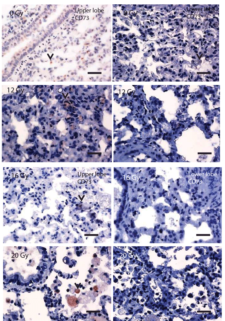
| Figure 4: Immunohistochemically stained micrographs for CD73 positive cells in control (0 Gy), 12 Gy, 16 Gy, and 20 Gy irradiated rats. The left four micrographs are taken from the upper lung lobe while the right four are taken from the lower lung lobe. Arrows show positive reaction in different cells. Scale bar: 35 μm at 400x. |