|
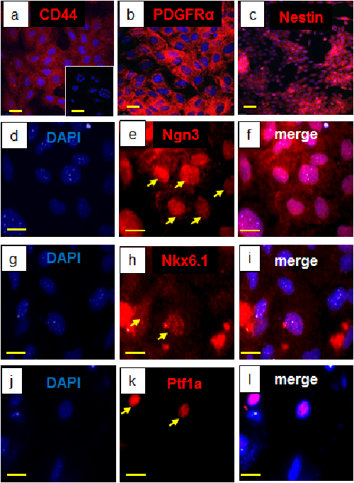
| Figure 1: Marker expression in MMMbz before and after differentiation. Before using the differentiation protocol, MMMbz cells expressed (a) CD44 (red), (b) PDGFRα (red), and (c) nestin (red). The inset in (a) shows isotype controls. After differentiation, the cells expressed (e) Ngn3, (h) Nkx6.1, and (k) Ptf1a. (d, g, j) DAPI staining. (f, i, l) Merged images. (a, b) bar = 25 μm. Inset in (a), bar = 100 μm. (c) bar = 100 μm. (d to l) bar = 10 μm. |