|
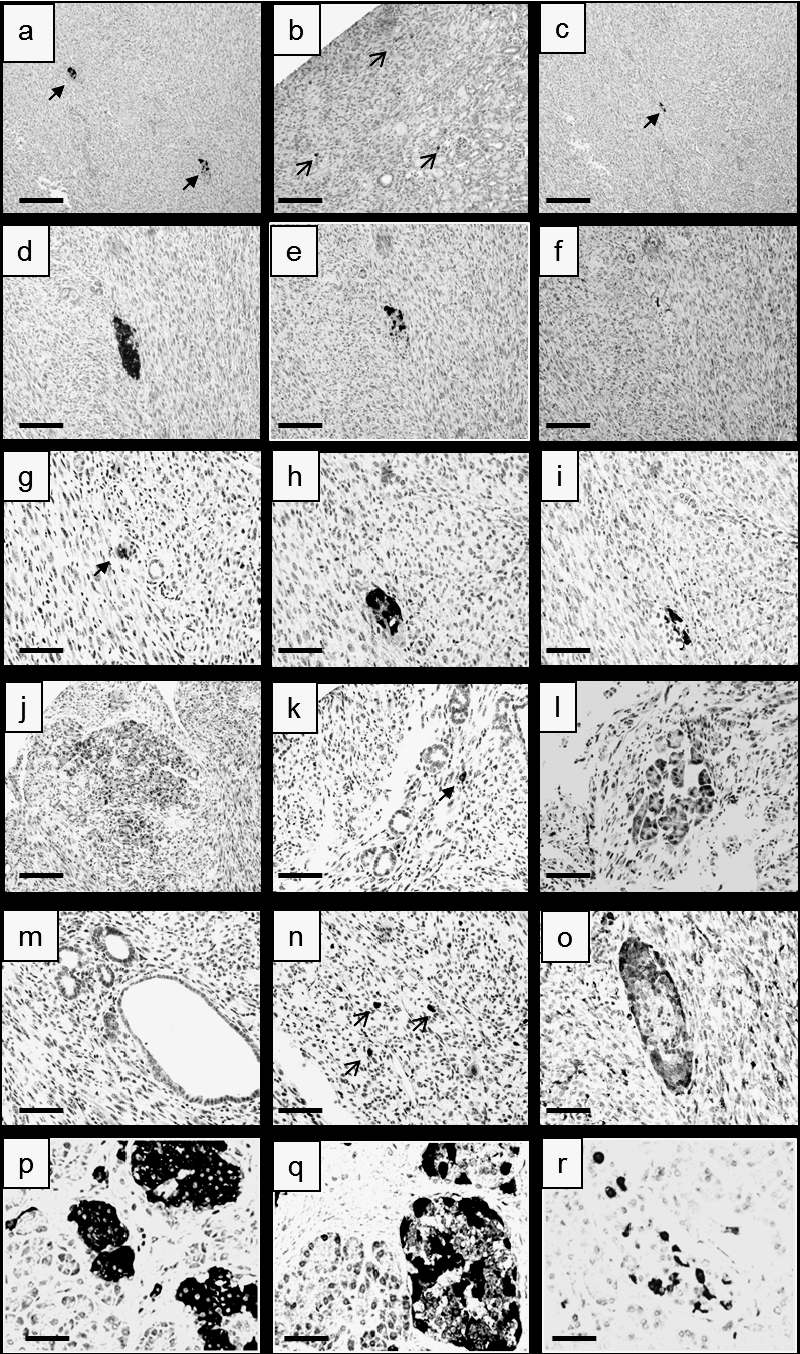
| Figure 5: Immunohistochemical analysis of the INS-GFP cells engrafted under the kidney capsule of SCID mice. (a) Islet-like clusters that expressed insulin (arrows). (b) Cells close to the kidney capsule that expressed GCG (arrows). (c) Clusters that expressed STT (arrow). (d) Higher magnification of islet-like clusters showed in “a”. (e) The same cluster that expressed GCG and (f) STT. (g) Different cluster that expressed insulin, (h) GCG, and (i) STT. (j) Acini-like structure. (k) Epithelial-like cells with insulin positive (black) cells (arrow). (l) Acini-like structure in a different field. (m) Cavities lined by epithelial-like cells. (n) Cells that expressed Ki67. (o) Islet-like cluster that co-expressed insulin (not shown) and Ki67 (black). (p) Normal mouse islet that express (p) insulin, (q) GCG, and (r) STT. (b, d, e, f, j, k, l) Bar = 250 μm. (g, h, i, m, n, o, p, q, r) Bar = 100 μm. (a, c) Bar = 400 μm. |