|
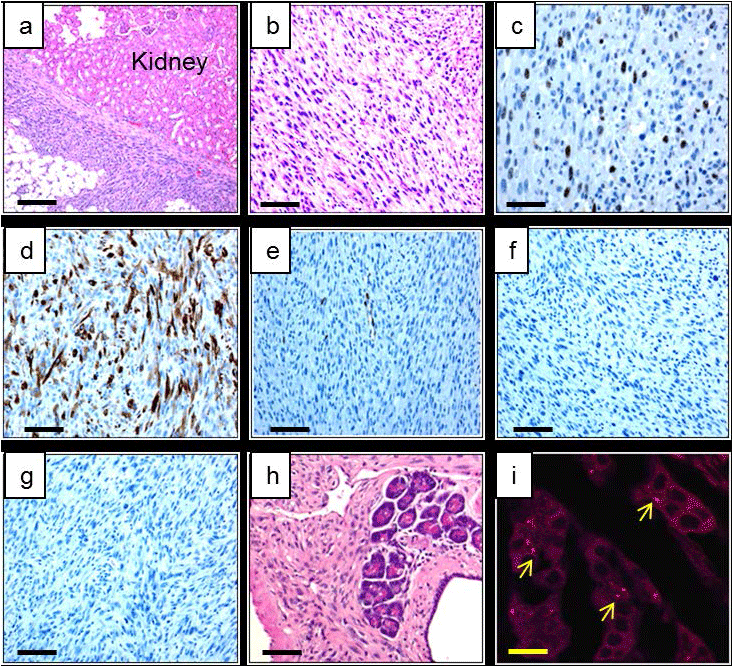
| Figure 6: Immunohistochemical analysis of the tumor developed in engrafted SCID mice. (a) Hematoxylin and Eosin (H&E) staining of the undifferentiated sarcoma close to the kidney capsule. (b) Higher magnification of the sarcoma stained with H&E. (c) Tumor cells stained for Ki67, (d) smooth muscle actin (SMA), (e) desmin, (f) AE1/AE3, and (g) S-100 protein. (h) Acini-like structures found within the tumor stained with H&E. (i) Higher magnification of these acini-like clusters stained also for anti-macro-H2A (Barr body). (a) Bar = 250. (b – h) Bar = 100 μm, (i) Bar = 25 μm. |