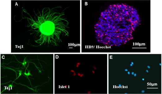
Immunofluorescence staining for various motor neurons markers: anti-Tuj1 (green, A) and anti-HB9 (red, B) in EBs derived from mESCs; anti-Tuj1 (green, C) and anti- Islet-1 (red, D) in dissociated MN cultures by higher magnification image. Hoechst (blue) was used to identify the nuclei. Scale bar: 50 μm and 100 μm.