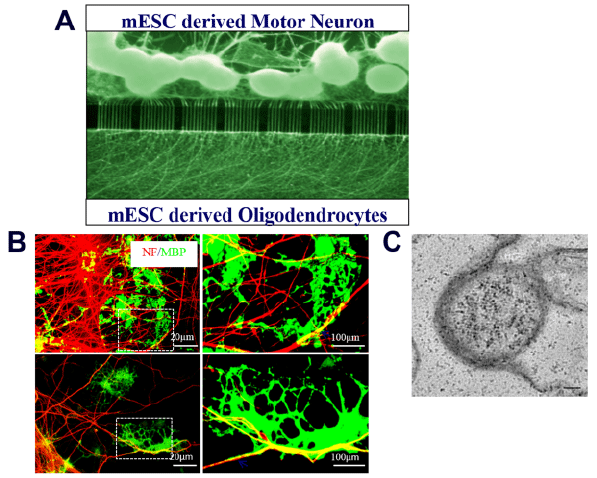
(A) Axons from mESCs derived MNs are guided by the microchannels and reach to the oligodendrocyte compartment. (B) Myelin sheaths (yellow colour) are defined as completely overlap between MBP+ oligodendrocyte processes (green) and NF+ axons (red) (Ba and Bc) and higher magnification image (Bb and Bd, enlarged image from Ba and Bc in dotted lines). (C) The electron microscope image of a myelinated axon fiber. Multi-layer of myelin around the axon fiber is observed. Scale bar: 20 μm and 100 μm.