|
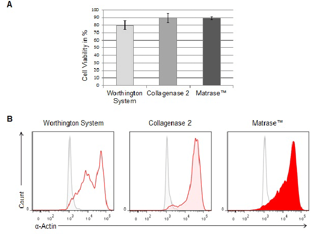
| Figure 2: Purity and viability of the isolated cell population comparing different isolation methods. (A) Cell viability was assessed immediately after isolation by trypan blue staining. Analysis was performed in three independent sets of experiments. Data represent the percentage of viable cells relative to total cell number. (B) Freshly isolated cells were fixed, permeabilized and labeled with a DyLight 594 fluorescent antibody against cardiac-specific α-actin. Flow cytometric analysis was performed to compare the purity of the isolated cardiomyocyte population. The gray histogram represents the unlabeled control. |